Immunohistochemistry; Is Used To Diagnose The Cancer
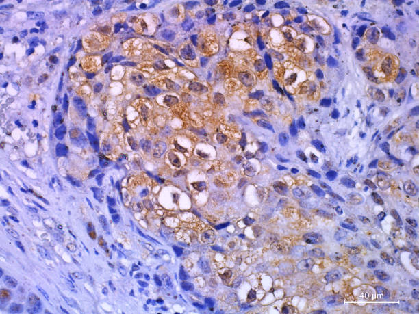
Immunohistochemistry (IHC) is a lab procedure that utilizes antigens associated to enzymes or fluorescent pigments to check for particular antigens in the biopsy cell sample. It can aid the care team inform one if the cells in the tissue specimen are normal, abnormal or malignant. Immunochemistry is depending upon the principle that antigens are very particular and only combine to particular antigens within cells. These antibodies can be proteins, peptides or DNA.
The antigens utilized for IHC staining can be monoclonal or polyclonal, based on the kind of target they are absorbed over and the method of identification they will be utilized for. Both kinds of antigens are helpful for identification in cells, however a single kind of antibody may not be sufficient to cover a vast range of targets. Species-matched antigens are diluted in PBS or in stopping buffer, further used on sections comprising the appropriate quantity of cells for a duration of time at room temperature. They are further rinsed thrice with PBS Tween-20.
According to Coherent Market Insights, the global Immunohistochemistry Market is predicted to reach US$ 1,463.6 million in 2022 and grow at a 6.6% CAGR from 2022 to 2030.
Secondary antigens, which are generally chromogenic and consist an enzyme conjugated to their crystallyzable fragment domain, are included to the section for staining. The enzymatic action of the secondary antigen necessity to be inhibited, such as by utilizing hydrogen peroxide or levamisole, to ignore confounding outcomes and destruction to epitopes. Immunohistochemistry detects cancer in an individual. As per CDC, around 1, 918,030 people in U.S were diagnosed with cancer in 2022 annually.
The section should be observed under the microscope for bg staining, which can be resulted by endogenous cell compounds. These consist proteins, other proteins and microbial DNA or RNA. IHC is a histopathological method that allows the location of cellular antibody within cells, which enables the identification of cell changes. It can be utilized to detect and localize the appearance of varied kind of varied particles, consisting enzymes, proteins, cytokines, and transcription reasons that are essential in biological procedures such as cell proliferation or death.
In IHC, antigens bind to the base of a cell sample and activate a color while they interact with the antibody. The dye consequently releases light that can be seen under a microscope, enabling the pathologist to detect the antibody present in the specimen. IHC can be conducted on formalin-fixed, paraffin-embedded and snap-frozen, cryosectioned cells. Formalin-fixed, paraffin-embedded segments are chosen for many routine histological uses however can be difficult for some kinds of antibodies.
In Immunohistochemistry, a primary antigen is combined to an epitope of a target protein and a supplementary antigen is included to the complex. Once the epitope-antigen binding occurs, a chemical substrate is included that reacts with the reporter particle and leads to a colored precipitate to create at the site of the whole antigen-antibody complex.


Comments
Post a Comment